Introduction
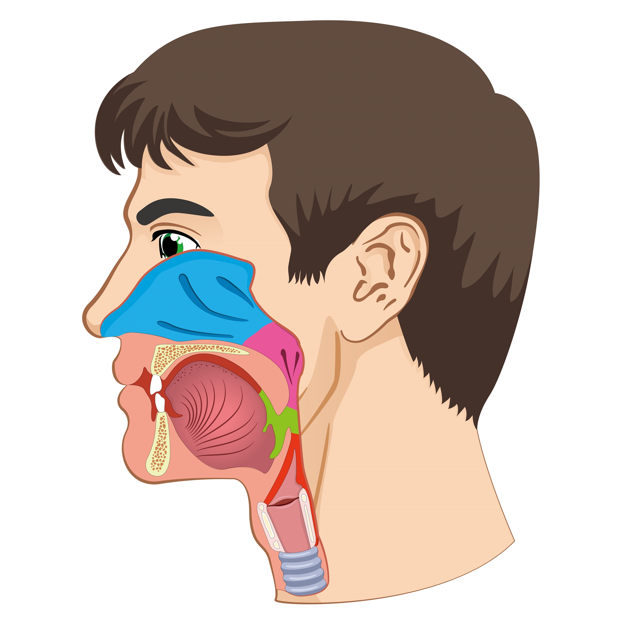
In Taiwan, hypopharyngeal cancer is ranked the third head and neck cancer with highest prevalence beneath oral and nasopharyngeal cancer. The occurrence of hypopharyngeal cancer is in close relation with smoking, alcohol and betel quid. 95% hypopharyngeal malignancies are squamous cell carcinoma. Patients diagnosed with hypopharyngeal cancer are typically men aged between 55-70 years old. Hypopharyngeal cancers are often named after their location, including pyriform sinus, posterior pharyngeal wall and postcricoid area. Most arise in the pyriform sinus (about 70%). About 70% of the patients had neck lymph node metastasis and 20% of them had distal metastasis (lung, bone, liver) at the time of diagnosis.
Etiology
The main risk factors associated with this disease are alcohol, betel quid, and tobacco use. Chronic irritation of the pharynx from gastroesophageal or laryngotracheal reflux of the gastric contents has also been associated with the development of tumors.
Symptoms
The symptom is not significant in early stage and the disease is often advanced at the time of diagnosis. If carefully asking about the history, we can find symptoms like sore throat, lump sensation, and otalgia. But the symptoms are just easily confused with common cold or chronic pharyngitis.
Symptoms of Hypopharyngeal Cancer include:
- Swollen lymph nodes in the neck
- Sore throat in one location that persists after treatment
- Pain that radiates from the throat to the ears
- Difficult or painful swallowing (often leads to malnutrition and weight loss because of a refusal to eat)
- Voice changes (late stage cancer)

Diagnosis
We can evaluate tumor location, cord movement via indirect mirror or fiberscope. Besides, neck palpation should be done for neck lymph node metastasis.
- Imaging studies
CT scan and MRI are used to visualize the primary tumor and regional lymph nodes prior to definitive treatment. Chest x-ray films and CT were necessary for lung metastasis. Liver echo and bone scan can detect liver and bone metastasis. The use of FDG-PET has been demonstrated to be useful in staging head and neck cancers.
- Laryngomicrosurgery:
Under general anesthesia and microscope, we can check the location of tumor and biopsy for tissue proof.
- Esophagoscope and bronchoscope
Esophagoscope can be used to assist in defining the inferior extent of the tumor and synchronous second primary esophageal tumors. Bronchoscopy is performed to rule out trachea involved.
Treatment
The treatments of hypopharyngeal cancers include surgical excision, radiotherapy, and concurrent chemo-radiotherapy. Earlier-stage cancers are treated with operation or radiotherapy alone. Combined surgery and postoperative radiotherapy or concurrent chemoradiotherapy has been one of the standard treatments for patients with advanced hypopharyngeal cancer. Surgery for advanced hypopharyngeal cancer may impair the swallowing and speech functions and, therefore, may have a negative impact on the patient’s quality of life. Organ preservation is achieved in patients with advanced cancer via concurrent chemoradiotherapy or neoadjuvant chemotherapy followed by radiotherapy or concurrent chemoradiotherapy, but not all patients with advanced hypopharyngeal cancer are indicated for aforementioned treatment modality.
Prevention
Most cases of hypopharyngeal cancers can be prevented by avoiding the factors that are known to increase the risk of these diseases, especially betel quid, tobacco, and alcohol.

